Heart Valve:
We learned a lot about heart valves over the past month.
The reason someone would need a heart valve replacement is something called regurgitation. This occurs when after blood is pumped one way, some leaks back into the heart the other way and can cause heart failure over time.
When someone needs heart valve replacement surgery, there are a couple routes they could take. The first type is mechanical replacement. This uses man made materials such as stainless steel or titanium. These are valves with opening and closing mechanisms that are inserted into the heart. These valves last the longest and are the most trustworthy.
The second option is biological. This is performed by using human or animal flesh for the replacement. These replacements last around 15 years and the patient would need to take blood thinners for life
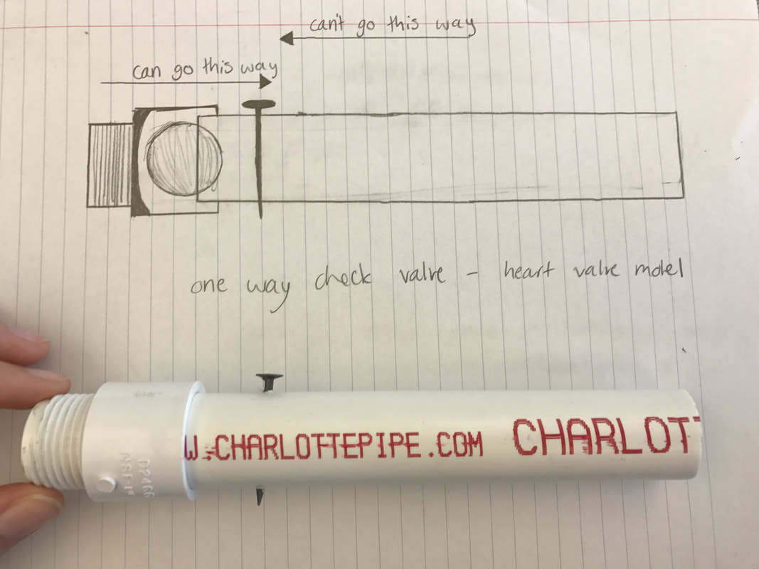
Our group decided to do a mechanical valve with a ball and cage design. When fluid is pumped one way, the ball allows it through and when it flows the other way, the ball plugs a hole and no fluid gets through. There are no elastic parts in this design, and if there were it would cause many problems and is a very risky factor to have present over time.
Here is the link to our padlet: padlet.com/saurban18/o518dopfcqn8
Notes:
- These tissue-paper thin membranes attached to the heart wall constantly open and close to regulate blood flow.
- There are two types of prosthetic valves used for replacement: tissue heart valves and mechanical heart valves. Both are designed to mimic the function of a natural, healthy heart valve.
- A mechanical valve is usually between 25-31 mm
- The tricuspid valve controls blood flow from the right atrium to the right ventricle.
- The pulmonary (pulmonic) valve controls blood flow from the right ventricle into the pulmonary artery that delivers blood to the lungs.
- The mitral valve controls blood flow from the left atrium to the left ventricle.
- The aortic valve controls blood flow from the left ventricle into the aorta.
- How do the heart valves function?
- As the heart muscle contracts and relaxes, the valves open and shut, letting blood flow into the ventricles and atria at alternate times. The following is a step-by-step illustration of how the valves function normally in the left ventricle:
- After the left ventricle contracts, the aortic valve closes and the mitral valve opens, to allow blood to flow from the left atrium into the left ventricle.
- As the left atrium contracts, more blood flows into the left ventricle.
- When the left ventricle contracts again, the mitral valve closes and the aortic valve opens, so blood flows into the aorta.
- As the heart muscle contracts and relaxes, the valves open and shut, letting blood flow into the ventricles and atria at alternate times. The following is a step-by-step illustration of how the valves function normally in the left ventricle:
- What is a heart valve disease?
- Heart valves can have one of two malfunctions:
- Regurgitation: The valve(s) does not close completely, causing the blood to flow backward instead of forward through the valve.
- Stenosis: The valve(s) opening becomes narrowed or does not form properly, inhibiting the flow of blood out of the ventricle or atria. The heart is forced to pump blood with increased force in order to move blood through the stiff (stenotic) valve(s).
- Heart valves can have both malfunctions at the same time (regurgitation and stenosis). When heart valves fail to open and close properly, the implications for the heart can be serious, possibly hampering the heart’s ability to pump blood adequately through the body. Heart valve problems are one cause of heart failure.
- Heart valves can have one of two malfunctions:
- ¾ inch PVC tubing
- ¾ inch male PVC slip adaptor
- ¾ inch bouncy ball
Reflection :
I enjoyed working on this project. I was interested in learning the actual physics behind the functions of the heart like how the valves open and close. We had a hard time designing the heart at first, because we focused on recreating the anatomy of a heart valve. After a couple tests, we decided on a more sturdy and permanent design. Our valve successfully blocked fluid from entering one side, while allowing it through the other, as it would need to in a heart. It would have to be shrunken and tweaked many times before entering the heart but our product performed the function it needed to consistently and without error.